 |
 |
|
Apert's & the Enigma of Chromosome No.10
Craniofacial Surgery: Tennessee Craniofacial Center Erlanger Health System Erlanger on Apert: Nat'l Org. for Rare Disorders: Apert's & Related Conditions Craniofacial Work at Columbia: Apert Syndrome: Faces-Cranio Disorders Clinical Variability in Patients: |
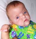
A CONGENITAL DEFECT FOR ONE IN 200,000 ... BUT WHAT IS APERT SYNDROME? From data in an article entitled "Clinical Assessment and Multispecialty Management of Apert Syndrome," Lawrence C. Kaplan, MD, published in Clinics in Plastic Surgery - Vol. 18, No. 2, April 1991. What Is Apert Syndrome? Major Features of Apert Syndrome
Possible Related Features of Apert Syndrome
Definition In a normal child, the skull is made up of several "plates" which remain loosely connected to one another, gradually growing together to form the adult skull. The Apert child's skull, by contrast, has a premature fusion of these plates, restricting brain growth, and causing increased pressure in the brain as it grows. This is known as craniosynostosis. Early surgery relieves the pressures by allowing the plates to be detached from one another. During this early surgery some "cranial remodeling" may be done to give the child a more normal appearance. The "retrusion" or hypoplasia of the midface is what could be described as a concave or dished in profile. As the skull grows, the upper and lower thirds of the face tend to grow at normal rates, but the middle third of the face grows slower, resulting in a more pronounced retrusion over time. A surgical procedure known as the LeFort III is used to correct this condition. The procedure is usually done after substantial growth is complete (preadolescence) and may be repeated as necessary. The LeFort procedure involves detaching the facial bones from mid eye to upper jaw and spacing this area out with bone grafts so that a proper alignment is made. The fusion of the fingers and toes along with the craniofacial problems mentioned above is what really separates Apert from other similar syndromes. This condition is called syndactyly. It always involves fusion of the soft tissues of the first, middle, and ring fingers, and often there is fusion of the bones themselves. Joint mobility is usually nonexistant past the first joint. The thumb may be fused into the hand, or may be free. Surgery is used to separate the fingers to obtain the highest degree of functionality, and may or may not ultimately result in five digits on each hand. It varies according to the degree of malformation. The feet and toes are affected similarly, but surgery is usually only recommended in cases where the ability to walk would be impared. Ideally, treatment of Apert begins at birth with the proper diagnosis, identification of the child's individual needs, and the proper facilities to administer what is needed. A multidisciplinary approach is used by physicans in the best arrangements. A craniofacial anomalies team may consist of a craniofacial surgeon, neurosurgeon, ENT, audiologist, speech pathologist, oral surgeon, psychologist, opthalmoligist, and an orthodontist. The team approach is used by these physicians to determine the best collaborative corrective plan for the deficiences of the child. |
|
|
APERT SYNDROME STORIES/SUPPORT Teeter's Page. Major site for Apert links and family-managed Apert ListServ. Apert Chat. Supplement to the Apert ListServ. An Excite community. Apert Support Group at Harvard. Andrea's Page. Personal account from Andrea Gartner, a college student with Apert Syndrome. Amy's Page. Personal account from Amy Esler, a girl with Apert Syndrome. Apert ListServ. An archive of ListServ postings on Apert Syndrome. ApertInfo: Story of Matthew Romero, in conjunction with Children's Hospital of Boston. Thrive Online: A med library article on Apert Syndrome. OTHER RESOURCES (These Links Will Open In New Browser Windows) Swedish Apert Syndrome Informat Medical College of Wisconsin: Apert Overview CCAKids PDF on Apert Syndrome FamilyVillage on Apert's Craniofacial Anomalies:Tooth Formation and Eruption in Patients WideSmiles: Apert Cleft Links
|
||
| eXalTerNet | TOP OF PAGE |